Just two posts ago I opined on the topic of whether the ordeal of the Achilles surgery, and more to the point, the ordeals of all the things runners have to cope with as a result of their habit, were worth the cost. I said then that the reward far outweighed the cost. Despite what I’ve been through in the last few days, I’ll stick to that. But wow, what a few days!
This week’s promise was to have been the start of my migration out of the splint and into the boot, most importantly off the crutches. But like a bolt out of left field, a freight train showed up and derailed that plan. Rather than hobbling around the house in the boot, I’m lying in a hospital bed getting pumped full of Drano and enjoying a fresh round of painkillers.
Say what, say you?
Let’s back up a bit, because by the time of waking to a public acknowledgement of my monogram on the radio, I already knew it wasn’t my day. Twenty-four hours earlier I’d awoken in moderate agony. I didn’t know it was merely moderate until I found true agony later that day, which gave me a better yardstick on which to measure such joy. The source of said agony wasn’t at all clear, but had I guessed my fate at that moment, the word kidney would have been involved. Stories I’d heard of how kidney stones felt like childbirth came to mind, but popping a pill and moving around a bit brought enough relief to believe there was a chance it was a fluke, if not a seriously ugly fluke, and that it was worth riding out. Most of the day, spent at my desk, passed with only mild aftereffects.
But that evening it was back, this time multiplied by two, with my shoulder now joining my kidney at first simply screaming, then expanding the scream exponentially to a veritable riot when I laid out on the couch for relief. No amount of Googling of “pain kidney shoulder” turned up anything rational or remotely plausible. Kidney and shoulder? One’s an organ, one’s a joint. Made no sense, but at least this time I figured out that staying upright made it (mostly) go away, which in my logic of the moment, hinted I didn’t need to race to the emergency room.
But it was a rough night, which led to being wide awake when that radio sprung to life saying, “GMC!” Call it the ultimate wake-up call, I suppose, because by this point it was clear this wasn’t a ride-it-out scenario. The only question was what to do first. I often say that you will have no way of knowing what the thing that finally kills you feels like, since you will, by definition, never have felt it before. Likewise here. This seemed pretty serious, but was it emergency-room-worthy in an era where everyone tells you that the ER is the most costly way to deliver medicine, and that you should use your primary care first? Still in the mode where sitting upright made it bearable, I opted for the economically advantageous choice, and headed for the office of Lady Doc.
Lady Doc, one of our heroes from blogs past, somehow carved a huge amount of time from what I presume was already a booked slate. Together we carefully covered as many bases as we could collectively think of, leading us through an odyssey of tests ranging from easy to successively harder questions. Pass the last one, you get an A. So who was the criminal element in this puzzle? To little fanfare, the theory of a bizarre nerve pinch left over from my lifting at the gym a few days back wasn’t realistically pursued. To my expectation, despite the oft-cited ‘pain-in-the-shoulder-means-you’re-having-a-heart-attack’, my ticker was preliminarily cleared (to be subsequently further cleared not only of blame, but collateral damage as well with another rollicking session on the echocardiogram with Tina the Tech of “Eighteen Again” fame; great to see you again, Tina!). To my surprise, the kidney was cleared; a great relief as I’d left the house half expecting to end up the target of stone-busting shock waves and the other half expecting to come home a kidney down.
Those discounted, that left a worrisome yet not unexpected, and altogether manageable scenario: a post-surgical blot clot, formally known in med-speak as an embolism, and in particular, based on the source of my agony, lung-based, or a pulmonary embolism. One more test, slide me into the CT scanner (another ten minutes of agony as was any time spent horizontally), and the verdict was rendered: Dude, you’re not going home tonight.
Cutting to the car crash, the final score wasn’t an embolism, but a cross country team of embolisms, the top runner being of considerable girth yet interestingly located on my pain-free side, entirely innocent of agony of the day. A collection of others joined in the fun – apparently it’s fairly common that when one is born, others follow, and they scattered themselves through the filter of my pulmonary arteries – but all had lodged themselves in places which still allowed blood to bypass. All but one. If not for that last one, I might have never known about this garden party, which might have been serene oblivion, or might have been fatal. Kind of hard to tell. But one of those buggers placed himself (I know it was a he, I just know it) down in the bottom back left corner – yes, right near the kidney – and effectively sealed off a very small bit-o-lung. That sole cubic centimeter took one for the team and sadly has likely been given a game misconduct, but in the process it largely created the agony entirely disproportionate to its size. Or so we think. One can never really be certain.
The science that eventually figured all this out is, as usual, quite intriguing. In the end, it was the CT scan, a technology that’s been around for quite a while, that found the smoking gun. While it uses different energy than an MRI (unfortunately with much more radiation, but it’s the price you pay), the basic idea is the same: take successive slice images of using frequencies that can discern different types of soft tissue. What was different from the MRI was that, perhaps due to the higher radiation, multiple scans from different angles were not taken. This would appear to limit the information revealed, but through some very crafty image processing, the system can create composite slice images at a ninety-degree angle from the images actually shot. While the raw slice images revealed the small dead spot – the pulmonary infarction in med-speak – It was these cross-cut composite images that gave clear pictures of a number of the clots. I was able to get a great education on this thanks to the significant time that Med School Mike, a third-year student at UMass Medical in Worcester, was willing to spend with me. His advisors should be assured that he’s got bedside manner nailed, not to mention plenty of expertise behind it. My hospital roomie must have been amused at what probably seemed like two kids playing with their toys, giddy at the images unfolding despite, or more accurately because of, their life-critical contents. (A collection of cool pics follows at the end of today’s extended tome.)
The images were so critical because the standard symptoms weren’t. The irony here is multi-level. The E. R. doctor made it quite clear that my fitness level, attained through running, obviously lessened the symptoms. As horrendous as the pain was (and as I’ve liked to point out to my caregivers, runners are used to pain, but this was PAIN), I displayed none of the other usual symptoms, which made the diagnosis that much harder. The best example is that most folks with clot-weakened lungs display obvious shortness of breath. I can’t tell you how many times I’ve been asked if I’ve been short of breath, and greeted with puzzled looks when I answer negatively, until the realization has dawned on the inquisitor that as a runner, there’s enough spare lung capacity that, especially when off the roads due to the Achilles repair, who’s going to notice that a chunk of it was (temporarily) disabled?
Moreover, that doc noted that said fitness might possibly have saved my life – which would be yet another time that running has done such a task. Yet running got me into this mess to begin with. After all, the Achilles tear was for a running injury, and even the need for repair was running-driven seeing as that the average Joe would have simply let the thing heal while hanging out on the couch for months.
At the end of the day, I was already down for the count, with at least a month before returning to the roads. This obviously adds another bump in the path coming back. But hey, bumps are nothing more than small hills, and I like hills, right?
Imaging Department
We start with a front-on shot of the lungs that they snap just before running the full CT scan. In this image, the left (which is really the right) shows a very sharp edge where the bottom of the lung hits the diaphragm. On the right side (which is really the left, from where the agony erupted), that corner is not so sharp. Aha, a clue!

We move on to a standard CT slice, this one being the bottommost slice. In the lower right (which is really the lower left, remember? Confused? Stick around!) there’s a fairly clear chunk of what we believe is infarction, or tissue damaged by being starved of blood. Note that the mass just above it (outside the red circle) is the diaphragm, not lung.

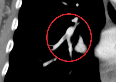
Now we move on to one of the clots. This one is from one of the standard CT slices. The white and gray shape is a pulmonary artery. The bright white is an artery full of blood, as it should be. The gray shadowy part is a blockage, without flowing blood. This one was the big one, we think, as it extended through several slices.

And finally, that same clot as viewed by the ninety-degree image-processed composite. In this one it’s really easy to see the pulmonary artery and the blockage inside.
Fascinating stuff, if you ask me. Enjoy.
Dude! That's very serious! But not a word on treatment or prognosis? You're killing us!
ReplyDeleteWorry not! I am on the mend! Your heartfelt concerns are appreciated and shall be answered, stay tuned!
ReplyDeleteThanks...